
Introduction
Here we will go over some additional information that will support your knowledge of both the cardiac cycle and the cardiovascular system as a whole.
The Electrocardiogram
The electrocardiogram, also known as the ECG, is an graphical representation of the electrical activity of the heart. It is produced by placing several electrodes around the body at specific points to measure electronic signals produced by the heart. It is an extremely useful tool for analysing and diagnosing various heart conditions.
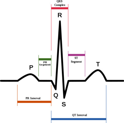
A typical ECG, as shown in the diagram above, consists of a P wave, a QRS complex (made up of the Q, R and S spikes) and a T wave.
The P wave is produced by atrial systole. A signal from the sinoatrial node is sent through the atria which depolarises them.
The QRS complex represents the rapid depolarisation of the ventricles. The waves appear in a complex way because of the different sizes of the right and left ventricles and the different times it takes for them to depolarise. The repolarisation of the atria also occurs during this time.
The repolarisation of the ventricles then produces the T wave.
Contraction of a Myocyte

During stage 0, depolarisation, sodium channels open releasing sodium ions into the cell. This is because, below a certain level of voltage, these voltage-gated channels open causing a sudden rise in voltage.
Phase 1, sodium channels close and chloride ions enter the cell, leading to Phase 2, plateau.
Calcium ions go into the cell via L-type calcium channels. This causes release of calcium via Ryanodine receptors, stored in the sarcoplasmic reticulum, which increases intracellular calcium concentration. This is what causes the plateau.
To ensure that repolarisation, phase 3 where voltage returns back to its negative level, potassium ions are released from the cell via multiple potassium channels until Phase 4 is reached.
Heart Abnormalities and Conditions
Listed below are several conditions of the heart that may be relevant to the cardiac cycle. We will provide a brief overview, so those wanting more detailed information should refer to the recommended textbooks
Angina:
Angina pectoris, commonly referred to as angina, is severe chest pain due to ischemia (lack of blood - usually in the coronary arteries) to the heart muscle.
Arrhythmias:
The term arrhythmia refers to a persistent change in the resting heart rate. A normal heart beats at around 75 beats per minute. Bradycardia is when the heart rate is too slow (less than 60 beats per minute) and tachycardia is when the heart rate is too fast (over 100 beats per minute). Arrhythmias can be caused by a variety of things, including drugs, electrolyte disturbances, abnormalities in the conduction pathways of the heart or just damage to the heart through trauma or another heart condition.
Coronary Artery Disease (CAD):
Coronary artery disease is the end-result of an accumulation of plaques in the coronary arteries that supply the heart muscle with blood and nutrients. CAD is the leading cause of death worldwide. Patients often may not show any symptoms for years before the first onset occurs (usually a sudden heart attack).
Myocardial Infarction:
A myocardial infarction, commonly known as a heart attack, is the death of heart cells due to the interruption of blood supply to them. A likely cause is due to the blockage of coronary arteries and the resulting ischemia.
Additional Reading
Medical Physiology, Walter F. Boron & Emile L. Boulpaep, Elsevier Health Sciences.
Anatomy & Physiology: The Unity of Form and Function, Kenneth S. Saladin, McGraw-Hill Education Europe.
Did you know..?
Dutch scientist Willem Einthoven recieved the Noble Prize for Medicine & Physiology in 1924 for his life's work developing the ECG.